Every year on March 3, conservationists, veterinarians, researchers, and wildlife advocates recognize World Wildlife Day. The day highlights the importance of protecting global biodiversity and encourages innovative approaches to safeguarding endangered species.
In the United States and beyond, one technology is making a measurable difference in conservation outcomes: portable ultrasound. Once associated primarily with hospitals and companion animal clinics, today’s compact, battery powered systems are supporting wildlife professionals in forests, wetlands, aquariums, rescue centers, and remote field sites.
Portable ultrasound in wildlife conservation provides real time, noninvasive imaging that allows specialists to make immediate, informed decisions while minimizing stress to animals. For organizations working with threatened and endangered species, that combination is invaluable.
Ultrasound imaging uses high frequency sound waves to visualize internal structures. It is safe, repeatable, and does not expose animals or handlers to radiation. These qualities make it particularly well suited to conservation work, where repeated monitoring and minimal disturbance are critical.
Captive breeding and managed reproduction programs are central to species recovery initiatives. Portable ultrasound allows biologists and veterinarians to:
• Track follicular development
• Confirm pregnancy
• Monitor embryo development
• Assess egg viability in reptiles
For oviparous reptiles, eggs appear as distinct, bright structures on ultrasound. In ovoviviparous species, embryos can be counted and monitored within individual sacs. This information helps determine optimal nutrition, enclosure requirements, and release timing.
The ability to perform these scans on site, without transporting animals to veterinary hospitals, reduces stress and improves overall welfare.
Portable ultrasound is also a powerful diagnostic tool for evaluating internal organ health. In zoos, sanctuaries, and field settings, it can assist in identifying:
• Gastrointestinal abnormalities
• Reproductive disease
• Bladder stones
• Cardiac enlargement or dysfunction
• Soft tissue masses
Because wildlife species often mask signs of illness, imaging plays a critical role in early detection. With portable systems, veterinarians and wildlife rehabilitators can perform immediate assessments and determine whether further treatment or intervention is necessary.
Aquatic conservation presents unique logistical challenges. Traditional imaging modalities are rarely practical near water or underwater. Portable ultrasound, however, has proven remarkably adaptable.
Researchers use ultrasound to examine fish, sharks, rays, sea turtles, and even coral structures. In certain aquatic environments, salinity and pressure conditions may enhance acoustic conduction, resulting in clear, detailed imaging.
These capabilities support health assessments of marine mammals, evaluation of reproductive status, and structural monitoring of coral reefs affected by environmental stressors.
Conservation rarely takes place in controlled clinical environments. Equipment must be lightweight, durable, and capable of operating on battery power for extended periods. Modern handheld and laptop based systems are designed to withstand demanding field conditions while delivering high quality imaging.
For wildlife professionals working in remote areas, portability is not a convenience; it is a necessity.
At ScanX Ultrasound, we have explored the potential of portable imaging in aquatic environments using systems such as the Vinno D6, the Apogee 1000 Lite, the Artemis, and the ScanX Air wireless probe.
The Vinno D6 paired with a linear transducer delivered impressive detail when imaging small aquatic species, including crustaceans in controlled tank settings. The Apogee 1000 Lite provided enhanced resolution for more advanced anatomical studies.
The Artemis system features a waterproof curvilinear transducer with a broad footprint, making it particularly well suited for scanning larger fish species.
Our ScanX Air wireless probe offers dual transducers, convex and linear, within a single handheld unit. Unlike many wireless systems, it maintains high resolution while connecting seamlessly to a tablet or smartphone. Although the probe itself is not fully waterproof, it can be used safely in aquatic settings with an appropriate protective barrier and coupling gel.
These systems have enabled detailed imaging of coral formations and marine life anatomy, demonstrating how portable ultrasound can support noninvasive marine conservation research.
In the United States, herpetologist Doug Wynn has utilized portable ultrasound technology to study endangered snake populations, including the Timber Rattlesnake, Eastern Massasauga Rattlesnake, and Plains Garter Snake.
These species are ovoviviparous, meaning they give birth to live young. On ultrasound, embryos are visible within individual sacs, allowing researchers to count developing offspring accurately. In one case, 27 embryos were identified in a single female.
The KX5600v system is highly effective for confirming pregnancy and identifying eggs. For more advanced imaging, the Apogee 1000 Lite provides additional detail, including the ability to visualize embryonic heartbeats using color Doppler technology. Monitoring blood flow and cardiac activity offers deeper insight into developmental health and reproductive success.
This type of information enhances conservation planning and long term species monitoring.
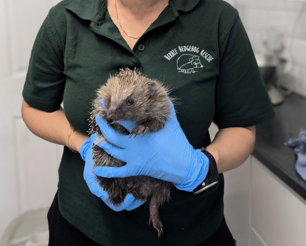
Portable ultrasound is equally transformative in small wildlife rescue settings. At Herbie Hedgehog Rescue, founded by Shweta Saikumar in the UK, the ScanX system has significantly improved intake and treatment protocols.
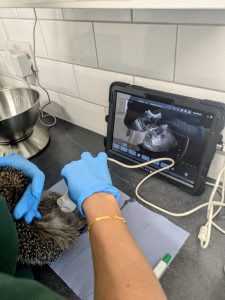
When female hedgehogs arrive at the rescue, ultrasound is used to confirm pregnancy status. This directly influences housing, nutritional planning, medication decisions, and release timing. Avoiding unnecessary transportation to veterinary clinics reduces stress and supports better recovery outcomes.
Beyond pregnancy detection, ultrasound assists in identifying uterine infections such as pyometra, bladder stones, gastrointestinal changes, and cardiac enlargement consistent with dilated cardiomyopathy. For nonprofit rescues operating on limited budgets, having in house imaging capability reduces costs while elevating standards of care.
Portable ultrasound in wildlife conservation is more than a convenient diagnostic tool. It represents a shift toward minimally invasive, real time, data driven wildlife management.
By reducing stress, delivering immediate results, and enabling repeat evaluations over time, portable systems support ethical and effective conservation practices. As artificial intelligence assisted image analysis continues to evolve, interpretation and measurement tools will further expand what is possible in field research.
On World Wildlife Day, we are reminded that conservation progress depends on innovation as much as passion. Portable ultrasound is helping wildlife professionals make smarter decisions, improve animal welfare, and protect vulnerable species across the United States and around the world.